Twelve patterns. Not twelve conditions.
The name on your diagnosis matters less than what is actually driving the pain, and two people with the same diagnosis often need opposite treatment. That is why five providers can all be competent and all be wrong at the same time.
What follows are twelve composite cases. Each one is built from the patterns we see most often, and each one shows the same four things: what the patient arrived with, what the measurements found, what we treated in what order, and where the numbers finished.
Read the one closest to your situation. If none of them fit, that does not mean we cannot help you. It means you should read Case 12 and then apply.
Case 01 · The patient who had back surgery and is still in pain
Pattern · Failed back surgery syndrome
Profile · Male, 58, retired construction project manager
What he arrived with
An L4-L5 fusion three years earlier. Six months of physical therapy before it and nine months after. Two epidural steroid injections. A second surgical opinion that recommended extending the fusion a level. Pain averaging 7 out of 10, worst in the morning and after sitting. He had been told the hardware was solid and the imaging looked good. Both of those things were true.
What the exam found
His primary driver was not the fused segment. Surface EMG showed the multifidus muscles on both sides of the fusion were barely firing, a shutdown pattern that persists long after the surgical reason for it is gone. His Dynamic Mobility Score was the lowest of the five, driven by almost no lumbar rotation and a compensating hip. qEEG showed a sensitized central pattern consistent with three years of continuous input. Driver classification: dyskinesis with a nociplastic overlay. Two categories, and the surgery had addressed neither.
What we treated, in what order
Pain Protocol first, two weeks, to bring the alarm down enough that he could tolerate loading. Then Retrain the Brain and Align the Structure together, using the Neubie to recruit muscle he could not voluntarily access, and graded exposure to the movements he had been avoiding for three years.
Where the numbers finished
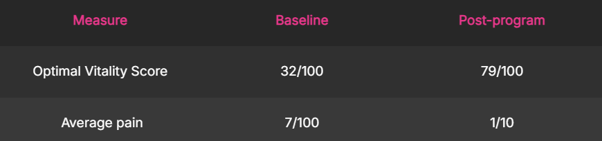
He was not a candidate for a third surgery. He was a candidate for the muscles nobody had turned back on.
Composite case example. Individual results vary.
Case 02 · The patient whose back pain has no explanation
Pattern · Chronic low back pain, six months or longer
Profile · Female, 41, dental hygienist
What she arrived with
Fourteen months of low back pain with no injury she could point to. An MRI showing mild disc degeneration at two levels, which four different providers described to her in four different tones of voice. Chiropractic care that helped for two days at a time. A prescription for a muscle relaxant she stopped taking because it made work impossible.
She had started to believe her spine was simply wearing out at 41.
What the exam found
The disc findings on her MRI are extremely common in people with no pain at all. In a study of more than 3,000 people without any back symptoms, disc degeneration was present in 37 percent of 20 year olds and 96 percent of 80 year olds. A scan alone cannot tell you what is driving your pain. What her measurements found was a right-dominant standing pattern from years of chairside work, almost no thoracic rotation, and a glute that switched off under load. Her Musculoskeletal Plasticity Score showed dense trigger point activity through the lumbar paraspinals and quadratus. Her Neuro Capacity Score was the strongest of the five, which was good news, because it meant the nervous system was not the primary problem.
Driver classification: dyskinesis, primary and almost isolated.
What we treated, in what order
Short Pain Protocol, one week. Then Align the Structure and Mobilize the Body as the main effort, with the work station itself rebuilt under Enhance the Environment, because a treated body returning to the same eight-hour posture is a treated body that relapses.
Where the numbers finished
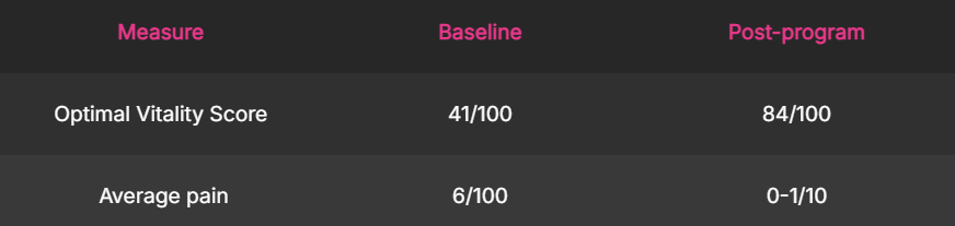
Her spine was not wearing out. Her right hip had been doing the work of her left for nine years.
Composite case example. Individual results vary.
Case 03 · The patient told the pain is "in your head"
Pattern · Fibromyalgia and widespread central sensitization
Profile · Female, 52, accountant
What she arrived with
Eleven years of pain in her neck, shoulders, hips and hands. A fibromyalgia diagnosis in year four. Normal bloodwork, repeatedly. Two rheumatology consults. Weekly flares that cost her two or three days each. A rotation through gabapentin, duloxetine and amitriptyline, all of which blunted the pain and flattened her thinking.
Somewhere in year six a physician told her it was in her head. She never went back.
What the exam found
He was wrong in the way that matters and accidentally close in the way that does not. The pain was not imagined. It was central, which is a different thing entirely and it is measurable. Her qEEG showed the slowed dominant rhythm and altered connectivity pattern that turns up consistently in widespread pain presentations.
Her heart rate variability was low and her resting sympathetic tone was high, meaning the alarm system was running continuously rather than switching on and off. Her Integrated Pain Score reflected pain in five regions with heavy sleep interference.
Driver classification: nociplastic, primary. The one category no injection or manual therapy can reach.
What we treated, in what order
Pain Protocol three weeks, longer than usual, because a sensitized system needs the alarm turned down before it can tolerate anything else. Then Retrain the Brain as the dominant pillar, including oculomotor pain desensitization and reprocessing, with sleep and autonomic work under Enhance the Environment running the entire time. Loading came last and came slowly.
Research supports this order. In a randomized trial of pain reprocessing therapy published in JAMA Psychiatry, 66 percent of patients were pain free or nearly pain free at four weeks compared with 20 percent on placebo, and a five-year follow-up found the difference held.
Where the numbers finished
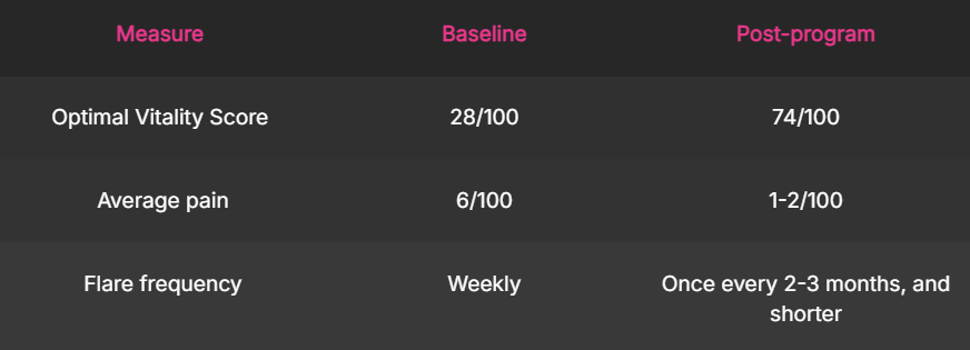
Eleven years of being told there was nothing to find, and it was on the scan the whole time. Nobody had run the right scan.
Composite case example. Individual results vary.
Case 04 · The patient whose headaches start in the neck
Pattern · Chronic neck pain and cervicogenic headache
Profile · Male, 36, software engineer
What she arrived with
Four years of daily neck stiffness and three to four headaches a week, always starting at the base of the skull and traveling behind the right eye. A migraine diagnosis and two preventive medications that did not change the frequency. Massage that helped for an afternoon. A cervical MRI reported as unremarkable.
He had stopped calling them headaches to people and started calling it his neck, which was closer to the truth than his chart was.
What the exam found
The pattern was cervicogenic, meaning the headache was referred from the upper cervical spine rather than generated as a primary migraine. PostureScreen measured significant forward head translation. His C1 assessment was asymmetric. Deep neck flexor endurance was a fraction of what it should have been, so the superficial muscles were holding his head up all day.
The oculomotor portion of his neuro screen was the finding that reframed the case. His eyes and neck were not cooperating, and every screen-based task was asking them to.
Driver classification: nociceptive with a dyskinesis driver, plus a neuro-oculomotor contribution.
What we treated, in what order
Pain Protocol one week. Then Align the Structure and Retrain the Brain together, deep neck flexor retraining paired with oculomotor and vestibular work, and Erchonia laser to the upper cervical region. His two monitors, their height, and the distance from his eyes were changed in week two under Enhance the Environment.
Where the numbers finished
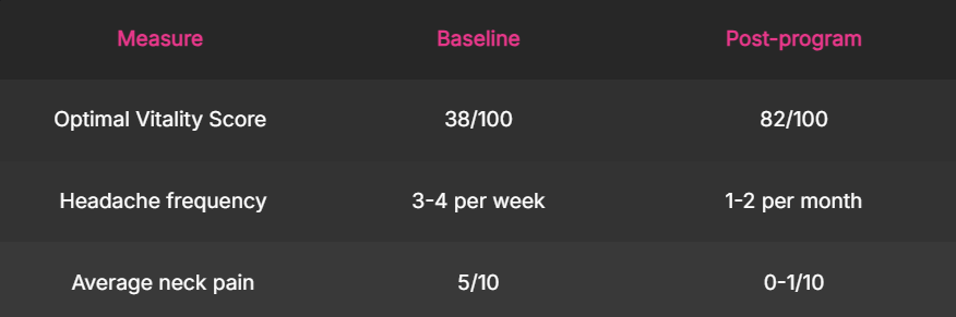
He did not have migraines. He had a neck that had been asked to hold his head six inches in front of his shoulders for eleven hours a day.
Composite case example. Individual results vary.
Case 05 · The patient with sciatica that will not quit
Pattern · Sciatica and radiculopathy
Profile · Female, 44, real estate agent
What she arrived with
Two years of right-sided pain from the buttock into the calf, with numbness across the top of the foot. An MRI showing an L5-S1 disc bulge. Three rounds of physical therapy aimed at the disc. One epidural injection with six weeks of relief. A surgical consult that put her in the "wait and see" category.
Long drives between showings made it worse, which meant her job made it worse.
What the exam found
Sciatica is a symptom description, not a diagnosis, and it has at least four common drivers that need different treatment. Her measurements found three of them stacked.
The disc bulge was real. So was significant hip dysfunction on the right with a deep gluteal that was not firing. So was multifidus shutdown at the two lowest segments. Sensory testing showed a true dermatomal pattern in the foot, confirming nerve involvement rather than referred pain.
Driver classification: neuropathic and dyskinesis together. The physical therapy had been aimed at the one driver least able to change.
What we treated, in what order
Pain Protocol two weeks with laser and neurological desensitization to settle the nerve. Then Align the Structure and Mobilize the Body, with hip and gluteal recruitment as the priority and multifidus retraining through the Neubie. The driver map dictated the order. Treating the hip before the nerve had calmed would have flared her.
Where the numbers finished
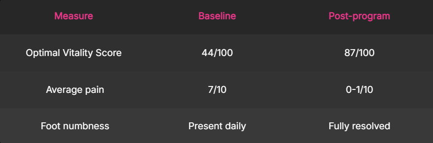
Three drivers, and two years of treatment aimed at one of them.
Composite case example. Individual results vary.
Case 06 · The patient whose new hip did not fix the pain
Pattern · Chronic hip pain, including pre and post replacement, FAI, and gluteal shutdown
Profile · Female, 63, retired school administrator
What she arrived with
A right total hip replacement fourteen months earlier. An excellent surgical result on imaging and a surgeon who was, correctly, satisfied. Groin and lateral hip pain that had never fully left, a limp she was told was habit, and increasing low back pain on the same side.
She had been discharged from post-surgical physical therapy as having met her goals.
What the exam found
Gait analysis showed a shortened stance time on the right and a trunk lean she was not aware of. The gluteus medius on the operative side had never come back, so the lateral hip was being stabilized by muscles that were not designed for it. Her lumbar spine was absorbing the difference, which explained the back pain that had appeared after the surgery rather than before.
Her Dynamic Mobility Score and Musculoskeletal Plasticity Score were both low. Her Neuro Capacity Score was intact.
Driver classification: dyskinesis, primary, with secondary nociceptive input from overloaded tissue.
What we treated, in what order
Short Pain Protocol, one week. Then Mobilize the Body and Align the Structure as the main effort, with gluteal recruitment through the Neubie, gait retraining with visual feedback, and progressive single-leg loading. Twelve weeks.
Where the numbers finished
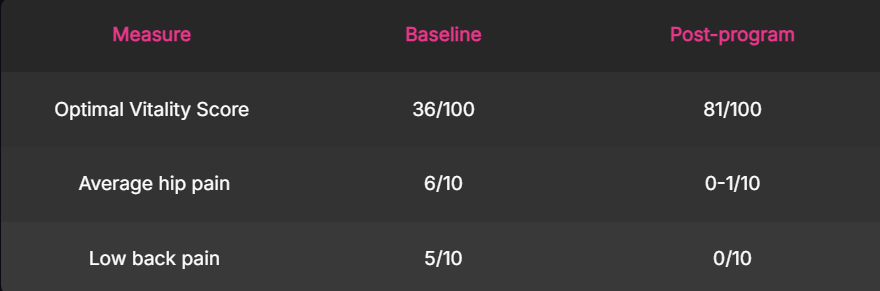
The joint was fine. The muscle that stabilizes it had been asleep for fourteen months and nobody had measured it.
Composite case example. Individual results vary.
Case 07 · The patient whose accident was two years ago
Pattern · Motor vehicle accident chronic pain, including whiplash, mild traumatic brain injury, and axial pain
Profile · Male, 47, insurance adjuster
What she arrived with
A rear-end collision at moderate speed twenty-six months earlier. Emergency department imaging that ruled out fracture. Four months of care that resolved most of the neck pain. What did not resolve: dizziness in busy visual environments, a persistent fog he described as thinking through water, sensitivity to fluorescent light, and mid-back pain that came back whenever he sat for more than an hour.
His case had closed. His symptoms had not.
What the exam found
The neck had largely healed. The vestibular and oculomotor systems had not. Smooth pursuit was broken into saccades, convergence was insufficient, and vestibulo-ocular reflex testing reproduced his dizziness directly. qEEG showed a pattern consistent with mild traumatic brain injury rather than a structural lesion, which is exactly why the emergency imaging was clean and correct.
His thoracic spine had almost no rotation, a guarding pattern set during the acute phase that had never been released.
Driver classification: neuropathic with central involvement, plus dyskinesis. Two years of treating the neck alone could not have reached either.
What we treated, in what order
Pain Protocol two weeks. Then Retrain the Brain as the dominant pillar, oculomotor and vestibular rehabilitation daily, with graded visual and vestibular exposure. Align the Structure and Mobilize the Body ran alongside to restore thoracic rotation. Light and screen conditions at his desk changed in week one.
Where the numbers finished
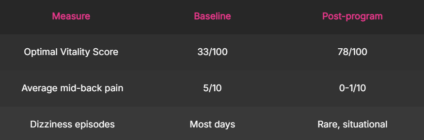
The collision hit his head as well as his neck. Only one of those got treated.
Composite case example. Individual results vary.
Case 08 · The patient told nerve damage is permanent
Pattern · Peripheral neuropathy, diabetic
Profile · Male, 66, retired postal supervisor
What she arrived with
Type 2 diabetes for fifteen years, reasonably controlled. Six years of burning and numbness in both feet, worse at night, with pain he rated 8 out of 10 in the evenings. Gabapentin at a dose that made him drowsy. Two balance-related near-falls in the previous year.
He had been told, more than once, that nerve damage does not reverse and the goal was slowing it down.
What the exam found
Sensory testing confirmed a symmetrical stocking distribution. What his chart did not contain was any measurement of the rest of the system. Balance testing showed heavy reliance on vision, meaning that when he closed his eyes or the room was dark, his nervous system had almost no information about where his feet were. That is why the near-falls happened at night.
His Neuro Capacity Score was low across sensorimotor and cerebellar domains. His Integrated Pain Score showed severe sleep interference, which matters clinically, because poor sleep raises pain sensitivity and creates a loop.
Driver classification: neuropathic, primary, with metabolic and autonomic contributors.
What we treated, in what order
Pain Protocol three weeks, laser and neurological desensitization, with sleep addressed immediately because the loop had to be broken first. Then Retrain the Brain focused on remapping sensory representation of the feet, with balance and proprioceptive loading, and metabolic and inflammatory factors addressed under Enhance the Environment in coordination with his physician.
Where the numbers finished
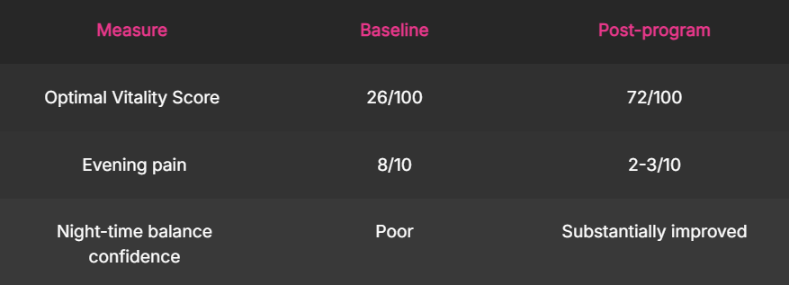
Nerves heal slowly. Nobody said they do not heal. What changed fastest was the brain's map of his feet, and that was never damaged in the first place.
Composite case example. Individual results vary.
Case 09 · The patient whose limb hurts to look at
Pattern · Complex regional pain syndrome, Type 1
Profile · Female, 39, hairstylist
What she arrived with
A wrist fracture four years earlier, cast for six weeks, healed without complication. Then swelling, color change, temperature difference, and pain out of all proportion to the injury. A CRPS diagnosis in month five. Two sympathetic nerve blocks with brief relief. Pain 8 out of 10 with the light touch of a sleeve reaching 9. She had not been able to hold scissors in that hand for three years.
She had been told nothing helps long term. She had also been told to use the hand more, without being told how.
What the exam found
The affected hand was distorted in her brain's body map. Two-point discrimination was markedly worse than the other side, she could not reliably identify which finger was being touched, and left-right judgment of hand images was slow and error-prone. Those are cortical findings, not tissue findings.
Autonomic measures showed the sympathetic overdrive that accounts for the temperature and color changes. Her Neuro Capacity Score was the lowest we measured across the five domains.
Driver classification: nociplastic with autonomic and neuropathic involvement. The blocks had targeted one part of a three-part problem.
What we treated, in what order
Pain Protocol three weeks with no direct handling of the limb, because in a sensitized system, touching the painful part first sets the whole program back. Graded motor imagery came first, then mirror work, then graded tactile discrimination, then movement. Retrain the Brain carried the case. Cortical remapping is where the current research points and it is the pillar the clinic is built around.
Progress was slower than any other case on this page. It was also the largest change in absolute terms.
Where the numbers finished
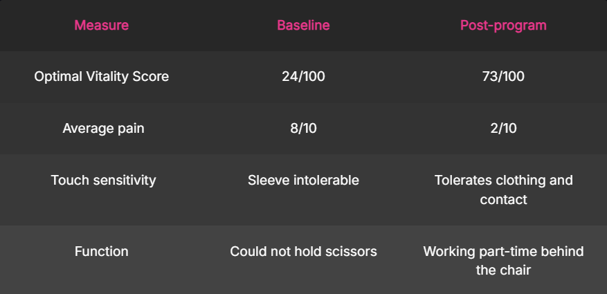
The hand was structurally fine four years ago. The map of it was not.
Composite case example. Individual results vary.
Case 10 · The patient whose pelvis was never examined
Pattern · Sacroiliac joint dysfunction and chronic pelvic pain
Profile · Female, 34, nurse, two children
What she arrived with
Five years of one-sided pain just below the belt line, dating to her second pregnancy. Pain with standing on one leg, rolling over in bed, and climbing stairs. A lumbar MRI that was clean, which she had been told meant nothing was wrong. Physical therapy directed at her low back. A pelvic floor referral she never completed because the waiting list ran nine months.
She had been given a maternity support belt in year two and told to keep using it.
What the exam found
The pain was not lumbar, which is why a lumbar MRI found nothing. Load transfer testing through the pelvis was positive on the symptomatic side. Her deep abdominal wall and pelvic floor were not coordinating with her breathing, so every step asked a joint with no muscular control to absorb the load on its own.
Her Musculoskeletal Plasticity Score showed dense adductor and gluteal trigger point activity, secondary to the instability rather than causing it. Her Functional Health Score showed a pelvic obliquity that had been read as leg length difference.
Driver classification: dyskinesis, primary, with nociceptive input from the joint itself.
What we treated, in what order
Pain Protocol one week. Then Align the Structure and Mobilize the Body as the main effort, deep abdominal and pelvic floor coordination retrained against the breath first, then load transfer, then single-leg and stair loading. The support belt came off in week four, deliberately, because a passive brace substitutes for the muscle you are trying to wake up.
Where the numbers finished
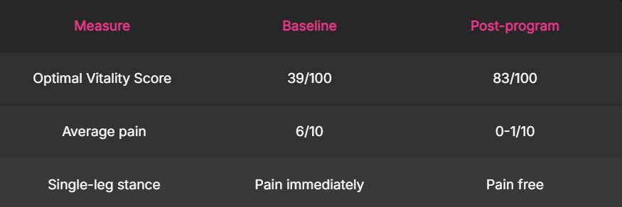
Five years of a clean lumbar MRI, and her lumbar spine was never the problem.
Composite case example. Individual results vary.
Case 11 · The patient on her fourth preventive medication
Pattern · Migraine and chronic tension headache
Profile · Female, 45, high school teacher
What she arrived with
Headache on eighteen to twenty days a month for six years, a mixture of migraine with visual aura and a daily band-like ache that was something else. Four preventive medications tried. Triptans that worked and that she was using often enough to be told she was at risk of a rebound pattern. Neurology imaging that was normal.
Her chart had one diagnosis. She had two problems.
What the exam found
Two patterns, running together. The migraine picture was neurological and her qEEG showed the cortical hyperexcitability pattern that is consistent with it. The daily band was cervicogenic and mechanical, coming from the upper cervical spine and the suboccipital muscles.
Autonomic measures showed low heart rate variability and high sympathetic tone, meaning very little physiological buffer against the things that set headaches off. Her sleep was fragmented. Her Neuro Capacity Score showed oculomotor findings that made screen work and fluorescent classroom light genuinely harder for her than for her colleagues.
Driver classification: neuropathic and central for the migraine, nociceptive and dyskinesis for the daily headache. Two drivers, two treatments, and a single diagnosis had been chasing one of them.
What we treated, in what order
Pain Protocol two weeks. Then Retrain the Brain and Align the Structure together, oculomotor and vestibular work paired with deep neck flexor and suboccipital retraining, and Erchonia laser to the upper cervical region. Autonomic and sleep work under Enhance the Environment ran the whole twelve weeks, because the buffer had to come back for anything else to hold. Medication decisions stayed with her prescribing physician throughout.
Where the numbers finished
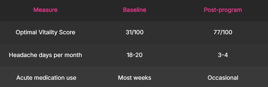
She was not failing four medications. Two of her problems were sharing one diagnosis.
Composite case example. Individual results vary.
Case 12 · The patient whose jaw pain was in her neck
Pattern · Temporomandibular and orofacial pain with cervical contribution
Profile · Male, 51, attorney
What she arrived with
Three years of jaw pain, worse on the left, with clicking, limited opening, and pain that spread into the temple and the ear. A night guard that reduced the morning soreness and changed nothing else. A dental evaluation confirming no decay or occlusal cause. Ear examinations that were normal, twice.
He had been told he clenched, which was true and was not the whole story.
What the exam found
Mandibular opening was limited and deviated to the left. The masseter and temporalis were dense with trigger point activity on that side. Then the finding that changed the plan: pressure on the upper cervical segments reproduced his temple and ear pain directly, and his deep neck flexors had almost no endurance. The head-forward position he held over documents all day was loading the jaw before he ever clenched.
His Neuro Capacity Score showed cranial nerve and oculomotor findings on the same side.
Driver classification: nociceptive with dyskinesis driving it from the cervical spine, plus an autonomic contributor from sustained stress load.
What we treated, in what order
Pain Protocol one week, laser and soft tissue work to the masticatory muscles for symptom relief only. Then Align the Structure and Retrain the Brain as the main effort, cervical and deep neck flexor retraining, jaw motor control, and oculomotor work. Desk height, document position and the two-screen setup changed in week two. The night guard stayed, as a passive protector, not as the treatment.
Where the numbers finished
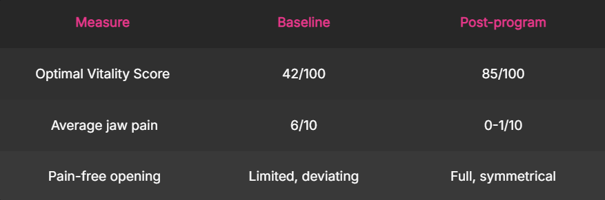
The jaw was the loudest part of the problem. It was not the source of it.
Composite case example. Individual results vary.
If your condition is not on this page
These twelve are the patterns we see most often. They are not the only patterns we treat. The condition name matters less than the driver underneath it, and we work from drivers. There are five: nociceptive, neuropathic, nociplastic, visceral, and dyskinesis. Most people arrive with two or three stacked, in an order nobody has mapped. Every case on this page is an example of that, not an exception to it. Apply, and on the Patient Interview we will tell you honestly whether your situation fits what we do. If it does not, we will say so, and we will point you toward what does.
What every one of these cases has in common
Not the diagnosis. Not the treatment. Every one of them had been seen by competent providers who measured part of the picture and treated what they measured. Nobody was negligent. The measurement was incomplete, so the treatment was aimed at the wrong driver, and it failed for a reason that was never the patient's fault. You cannot treat what you have not measured. That is the whole argument.
Composite case examples throughout. Individual results vary. These cases illustrate patterns we treat and are not representations of any specific individual patient.
Dr. Mark T. Wade, DC, DrPH · The Posture Clinic, a chiropractic practice · 6425 53rd St N Ste 6, Pinellas Park, FL 33781 · 727-233-5268
Sources cited in these cases
- Brinjikji W, et al. Systematic literature review of imaging features of spinal degeneration in asymptomatic populations. AJNR Am J Neuroradiol. 2015;36(4):811-816. https://www.ajnr.org/content/36/4/811
-
Brinjikji W, et al. MRI findings of disc degeneration are more prevalent in adults with low back pain than in asymptomatic controls. AJNR Am J Neuroradiol. 2015;36(12):2394-2399. https://pmc.ncbi.nlm.nih.gov/articles/PMC7964277/
-
Ashar YK, et al. Effect of pain reprocessing therapy vs placebo and usual care for patients with chronic back pain. JAMA Psychiatry. 2022;79(1):13-23. https://doi.org/10.1001/jamapsychiatry.2021.2669
- Ashar YK, et al. Pain reprocessing therapy for chronic back pain: five-year follow-up. JAMA Psychiatry. 2025;82(10):1049-1051. https://doi.org/10.1001/jamapsychiatry.2025.1844

(727) 233-5268
Clinic@PostureClinic.com
6425 53rd St N Ste 6, Pinellas Park, FL 33781
We don't manage chronic pain. We eradicate it.
The Posture Clinic, a chiropractic practice.
© The Posture Clinic 2026. All rights reserved.